Topic
Anatomy
Layer-by-layer facial anatomy, injection planes and the named vasculature.
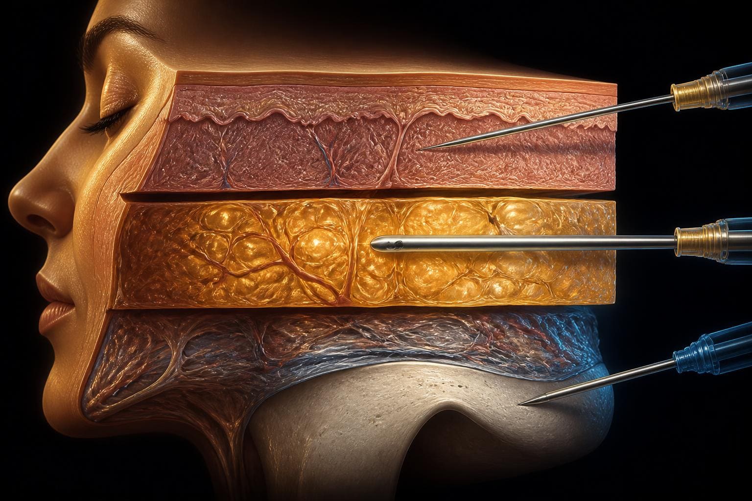
The Three-Layer Filler Model: Depth as the Anatomy of Injection
Every clinical discipline works with the anatomy its tools allow it to see. The surgeon confirms the course of a vessel under direct vision; the radiologist images the structure before anything is done. The aesthetic injector has neither — the needle works blind, and no amount of memorised anatomical variation can be verified at the moment of injection.
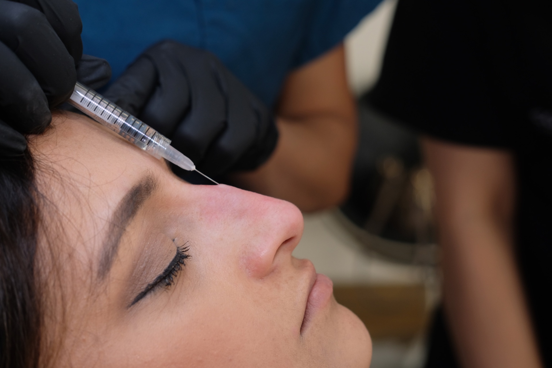
The Role of Injection Anatomy in Reducing Complications in Dermal Filler Practice
Vascular complications from dermal fillers are uncommon but devastating when they occur. The single most useful mental model for the non-surgical injector is not a map of named arteries — it is a map of depth. The vessels that matter are stratified, layer by layer, and the tool in your hand has to match the layer it is in.

Vascular Complications in Aesthetic Medicine: Prevention, Recognition and Management
Intra-arterial injection of dermal filler is the single most consequential complication in non-surgical aesthetic medicine. Although rare per millilitre injected, it carries the potential for skin necrosis, scarring and — in the case of retrograde embolisation to the ophthalmic circulation — irreversible blindness (Beleznay et al., 2019; Goodman et al., 2020). The window between event and irreversible tissue loss is short, and outcome is determined almost entirely by whether a written, rehearsed emergency protocol is executed within hours, not days.
Other topics